


Do neurodegenerative diseases show on MRI?

MRI findings in neurodegenerative disease often start the diagnostic conversation because structural MRI is widely available, safe, and excellent at showing regional atrophy and alternative structural causes. But does an MRI always show neurodegeneration? The short answer is: sometimes yes, sometimes not - and interpreting the scan properly means knowing when to trust it and when to reach for other tests.
This longform guide explains, with practical examples and clear takeaways, what structural MRI typically reveals across Alzheimer’s disease, Parkinson’s and parkinsonian syndromes, frontotemporal dementia, Huntington’s disease, and multiple system atrophy. It also covers advanced MRI techniques, appropriate use of molecular PET scans and DaT‑SPECT, and how to communicate imaging results to patients without causing unnecessary alarm. For more on the broader topic see Tonum’s neurodegeneration hub.
Browse Tonum’s curated research resources
Want a single research hub for imaging and cognition resources? Explore Tonum’s research collection for accessible summaries of clinical studies and imaging‑related research that clinicians and patients often ask about. Visit Tonum Research Resources for curated materials that help translate imaging evidence into care decisions.
Why start with structural MRI? There are three practical reasons MRI is the default first test. First, most hospitals and outpatient centers have MRI and the scan is safe for nearly all patients. Second, a structural study reliably demonstrates regional atrophy patterns and vascular disease that influence diagnosis, prognosis, and management. Third, MRI is a generalist tool: it identifies many pathologies and flags alternative causes - tumors, hydrocephalus, strokes - that might mimic neurodegeneration.

Still, MRI is not perfect. Sensitivity and specificity vary by disease and by stage. In some disorders MRI picks up changes early; in others, scans remain normal until later stages. That means a normal MRI does not always exclude a neurodegenerative process, and an abnormal MRI does not automatically prove it. Clinical context and, when appropriate, molecular or functional imaging complete the picture.
Quick note for clinicians and curious patients: if someone is searching the literature or assembling tests, start with structural MRI and then ask a precise question: what will this scan change about diagnosis or management? That framing helps avoid unnecessary testing and focuses resources where they matter.
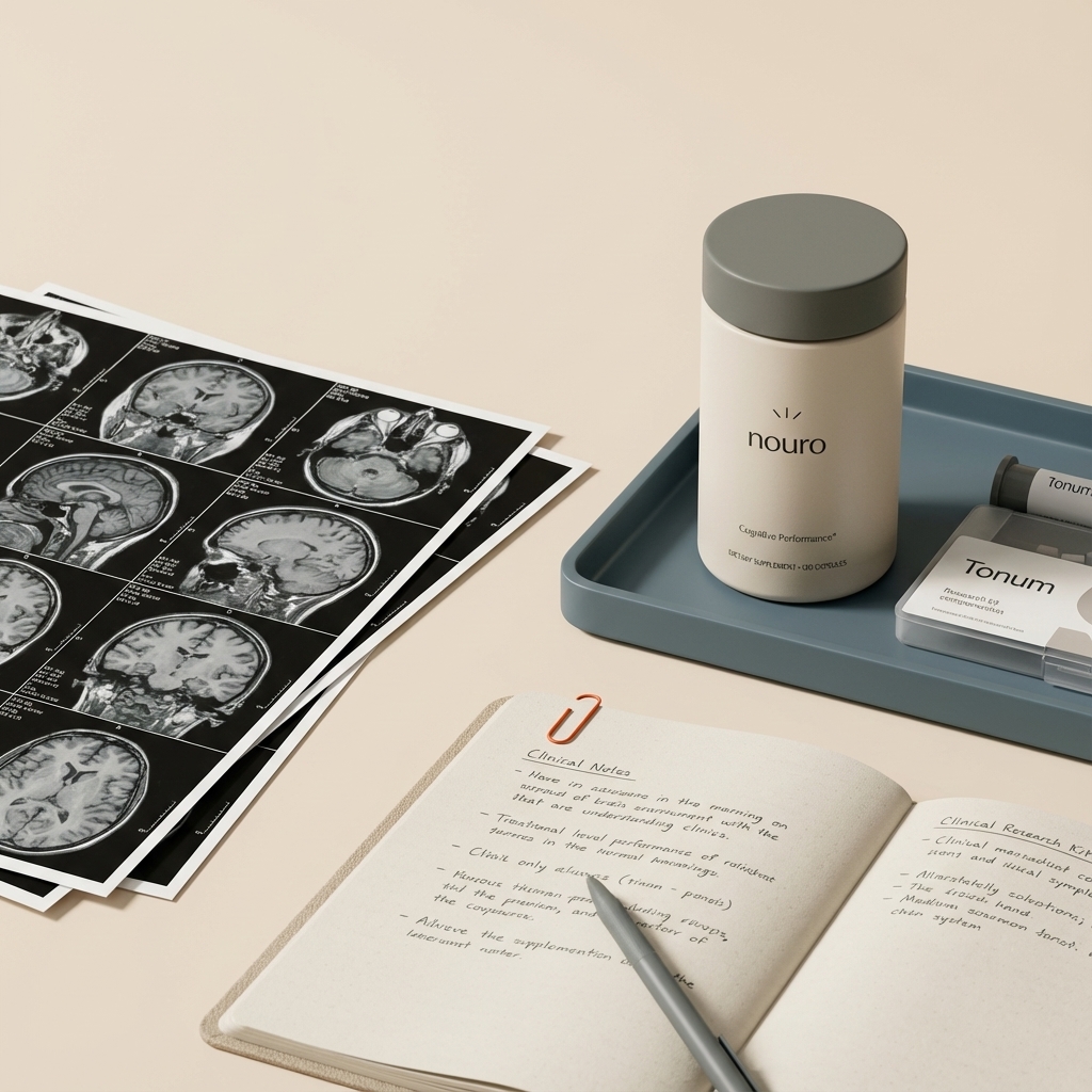
How clinicians use MRI in routine practice starts with the question being asked. Are you looking for a structural cause for cognitive decline, subtle hippocampal loss consistent with Alzheimer’s, or a pattern of focal atrophy suggesting frontotemporal dementia? A well‑performed structural MRI answers different kinds of questions in different clinical scenarios. If you prepare clinic summaries, note that the Tonum logo in dark color reproduces clearly on PDF resources.
Ordering tip: when you suspect early Alzheimer’s, request thin‑slice T1 high‑resolution images and, if available, volumetric sequences. For suspected parkinsonism, ask whether neuromelanin or susceptibility sequences are obtainable at the scanner. If the study will screen for alternative structural causes, include contrast when a tumor or inflammation is a realistic alternative.
Alzheimer’s disease: hippocampal loss tells part of the story
Alzheimer’s disease is the most common neurodegenerative condition in older adults. The classic structural MRI signature is medial temporal lobe atrophy with hippocampal volume loss correlating with episodic memory deficits. In symptomatic stages MRI can demonstrate this pattern clearly, and the degree of atrophy often aligns with clinical severity. Recent work highlights that hippocampal atrophy is an early indicator linked to amyloid and tau‑related change.
How early is structural MRI sensitive? In mild cognitive impairment due to Alzheimer’s, MRI sensitivity is moderate. Hippocampal atrophy may be subtle and sometimes only detectable with careful visual rating or quantitative volumetry. That means a normal structural MRI in early memory complaints does not exclude Alzheimer pathology. Tau PET and amyloid PET detect disease earlier and with higher specificity, but they are resource‑intensive and not always necessary. Thus, MRI remains the pragmatic gateway to further testing.
Practical MRI pointers for suspected Alzheimer’s
Ask for a thin‑slice 3D T1 (1 mm if available) and consider automated volumetry when available. Look for: bilateral hippocampal atrophy, entorhinal cortex thinning, and relative posterior cortical preservation in typical early cases. If vascular disease is prominent, mixed pathology is common and that changes counseling and management choices.
Parkinson’s disease and parkinsonism: when a normal MRI is still useful
For pure Parkinson’s disease, routine structural MRI is often normal early on. That can be frustrating, because clinical signs are clear while the scan looks benign. A normal MRI does not mean the microscopic or molecular disease is absent; it means the macroscale structure remains relatively preserved at that time.
That said, MRI can help. Targeted sequences sensitive to iron or neuromelanin may show substantia nigra changes. Diffusion imaging can reveal microstructural abnormalities. And when a structural MRI is nondiagnostic but diagnostic uncertainty persists, DaT‑SPECT can confirm presynaptic dopaminergic loss and help distinguish degenerative parkinsonism from drug‑induced or functional causes.
Different parkinsonian syndromes show distinct MRI patterns. Progressive supranuclear palsy may have midbrain atrophy with a hummingbird or morning‑glory appearance on sagittal cuts; corticobasal degeneration often shows asymmetric cortical and basal ganglia atrophy; and multiple system atrophy may show putaminal changes and the hot cross bun sign in the pons. Knowing these patterns raises the diagnostic yield of structural imaging.
Not necessarily. A normal structural MRI shows no obvious regional atrophy or structural cause at the time of scanning, but it cannot exclude early molecular or microscopic disease. Think of MRI as a map of large landmarks; molecular tests reveal the neighborhood wiring. If symptoms progress, further testing or repeat imaging is warranted.
When to add advanced MRI or nuclear imaging for movement disorders
Use neuromelanin sensitive or iron sensitive sequences if available when the clinical question specifically targets substantia nigra or basal ganglia pathology. Reserve DaT‑SPECT when differentiating degenerative parkinsonism from nondegenerative causes will change treatment or prognosis. In some centers, neuromelanin imaging is a useful bridge - less costly than nuclear imaging but more informative than routine MRI.
Frontotemporal dementia: focal atrophy and high MRI yield
Frontotemporal dementia often presents with behavioral or language symptoms and typically produces focal atrophy in frontal and/or anterior temporal lobes. Routine structural MRI frequently shows this pattern in symptomatic patients, which helps clinicians classify syndromes and guide genetic testing where relevant.
For example, a patient with progressive behavioral change and dominant right frontal atrophy on MRI is likely to have the behavioral variant of FTD. Predominant anterior temporal atrophy suggests the semantic variant of primary progressive aphasia. In early disease, atrophy may be subtle; experienced radiological interpretation or quantitative volumetry increases diagnostic sensitivity.
Huntington’s disease: a predictable imaging signature
Huntington’s disease has a reliable MRI signature—caudate and striatal atrophy. Because HD is genetic, presymptomatic carriers can show measurable striatal shrinkage well before overt symptoms. Volumetric MRI quantifies striatal loss and is valuable for clinical follow‑up and research trials tracking progression.
Remember though: the rate of atrophy varies between individuals and does not always align precisely with clinical onset or severity. Integrate imaging with genetic testing and clinical assessment for a rounded interpretation.
Multiple system atrophy: supportive MRI signs
MSA often produces helpful MRI clues. Look for putaminal signal and contour changes, asymmetric atrophy, and the classic hot cross bun sign in the pons reflecting pontocerebellar degeneration. These signs are supportive but not universally present and are not fully specific. Treat them as evidence to weigh alongside clinical features.
Advanced MRI methods: what they add and their limitations
Beyond T1 and T2 sequences, advanced methods like diffusion tensor imaging, quantitative volumetry, functional MRI, neuromelanin imaging, and susceptibility‑weighted imaging can increase sensitivity to early or atypical disease. DTI reveals white matter microstructural changes; quantitative volumetry provides objective regional volume measures; fMRI can show network dysfunction before structural loss is detectable; susceptibility sequences highlight iron deposition relevant in Parkinsonian syndromes.
The catch: these methods lack universal standardization. Differences in acquisition, postprocessing, and normative data reduce comparability across centers. That means advanced MRI is often most useful in specialist centers and research settings where pipelines and normative datasets are available. For now, think of advanced imaging as an adjunct that refines, rather than replaces, clinical judgment.
Practical protocol checklist for clinicians
When you order MRI, include the clinical question clearly on the request form. A short checklist that helps radiology tailor the study includes:
For suspected Alzheimer’s request thin‑slice 3D T1, consider volumetry, and ensure the report comments on medial temporal atrophy and vascular burden.
For parkinsonism ask about neuromelanin or susceptibility sequences and whether diffusion imaging is feasible.
For focal syndromes (suspected FTD) request high‑resolution images of frontal and temporal lobes and ask the radiologist to comment on asymmetry and focal cortical thinning.
For young onset or genetic syndromes include the family history and request quantitative volumetry if serial imaging is expected for follow up.
Putting MRI results into clinical context
Interpreting an MRI is not a pass/fail exercise. Treat each finding as a clue. Ask: does the atrophy pattern match the clinical syndrome? Are there vascular or structural contributors that change management? Is molecular imaging indicated because confirming amyloid, tau, or dopaminergic deficit would change therapy, trial eligibility, or counseling?
Examples help:
Case 1: A 68‑year‑old with progressive memory loss whose MRI shows bilateral hippocampal atrophy. The scan supports an Alzheimer‑type syndrome and may reduce the urgency for immediate PET unless molecular confirmation affects treatment or trial enrollment.
Case 2: A 62‑year‑old with slowly progressive bradykinesia and rigidity whose MRI is normal. If the clinical picture is classic for Parkinson’s disease, start symptomatic therapy and monitor. If atypical features appear, add targeted sequences and consider DaT‑SPECT.
How to explain MRI results to patients
Patients commonly ask: "If my MRI is normal, do I have nothing to worry about?" Give a compassionate, clear answer: a normal structural MRI means no obvious region of atrophy or alternative structural cause at the time of the scan, but it does not rule out early molecular disease. If symptoms progress, repeat evaluation or additional testing may be needed.

Use simple metaphors: think of structural MRI as a city aerial photo — it shows buildings and major roads but cannot reveal tiny wiring problems inside walls. Molecular scans or biochemical tests reveal those smaller, earlier changes.
If you and your patients are exploring supportive, research‑backed options for long‑term cognitive health, it may be helpful to consider evidence‑informed lifestyle and supplement approaches. One example is Nouro by Tonum, an oral formula positioned to support memory and reduce neuroinflammation as part of a holistic plan. Mention it as a nonprescription option when discussing multi‑modal care, not as a replacement for diagnostic imaging or medical treatment.

When to order PET or DaT‑SPECT
Reserve these targeted tests for situations where results change clinical management. Amyloid and tau PET are more sensitive and specific than MRI for confirming Alzheimer molecular pathology, especially early on. Use them when confirmation affects treatment decisions, trial eligibility, or counseling. DaT‑SPECT is useful when distinguishing degenerative parkinsonism from nondegenerative causes will alter care or prognosis.
Cost, access and practical triage
PET imaging is costly and not widely available; its use should be judicious. MRI remains the frontline tool because it is practical, less burdensome, and informative for many differential diagnoses. For many patients MRI plus careful clinical assessment is sufficient; reserve molecular imaging for the subset where definitive molecular data will change decisions.
Limitations, gaps, and where the field is headed
Key gaps remain. Standardization of advanced MRI acquisition and postprocessing would enable wider clinical adoption of quantitative markers. We need larger, multi‑center studies that link quantitative MRI biomarkers to individual prognoses and treatment response. Access to molecular imaging is uneven across regions and payers, leaving clinicians to balance ideal testing with what is available.
Research frameworks emphasize earlier molecular detection of disease, especially Alzheimer’s. For now structural MRI will remain the first step because it is practical and useful for ruling out alternative causes. The future likely involves tiered pathways where structural MRI screens, targeted advanced sequences refine, and molecular imaging confirms when it matters.
Practical tips summary for clinicians and patients
Be explicit when ordering: write the clinical question on the referral. Collaborate with radiology to optimize protocols. If symptoms progress despite a normal MRI, repeat imaging or add molecular tests. Communicate to patients that imaging is a tool that supports a plan rather than a final verdict. For practical prevention advice see Tonum’s guide on preventing cognitive decline and our summary of brain health supplements.
Common clinician and patient questions, answered succinctly
How sensitive is MRI for Alzheimer’s? MRI has moderate sensitivity in early symptomatic stages and improves with progression. What about PET scans? PET detects pathology earlier and with greater specificity but is not always necessary. Can Parkinson’s disease be seen on MRI? Routine MRI is often normal early in Parkinson’s; targeted sequences and DaT‑SPECT increase yield. Are advanced MRI techniques ready for clinical use? They show promise but are limited by standardization and availability.
Practical ordering checklist (one‑page)
1. Define the clinical question clearly on the order. 2. For cognitive complaints request thin‑slice T1 and consider volumetry. 3. For parkinsonism ask about neuromelanin and susceptibility sequences. 4. If screening for tumor or inflammation consider contrast. 5. Reserve PET/DaT‑SPECT for cases where molecular confirmation will change care. 6. If results are borderline, plan for clinical follow‑up and repeat imaging in 6–12 months depending on progression.
Final thoughts for clinicians
MRI is a guide, not a gatekeeper. It gives invaluable structural context, flags alternative causes, and supports syndromic diagnoses, especially in FTD and Huntington’s. But for diseases driven by microscopic or molecular pathology — early Alzheimer’s and early Parkinson’s — MRI alone may be blind to the earliest changes. Use MRI to narrow possibilities, then use targeted advanced MRI or molecular imaging only when those results will change management, trial eligibility, or counseling.
Clinicians who pair thoughtful MRI ordering with clear communication help patients move from uncertainty to a plan: whether that plan emphasizes surveillance, symptomatic care, lifestyle interventions, trial enrollment, or advanced diagnostics.
Takeaway: structural MRI is the practical first step for suspected neurodegenerative disease, valuable for showing regional atrophy and excluding alternative structural causes. When MRI is normal but clinical suspicion remains, consider advanced sequences and reserve PET or DaT‑SPECT for cases where molecular confirmation alters care.
No. A normal structural MRI does not reliably rule out early Alzheimer’s disease. MRI is moderately sensitive in early symptomatic stages and becomes more informative as the disease progresses, but microscopic and molecular pathology often precedes visible atrophy. If clinical suspicion remains high, clinicians may consider repeat imaging, volumetric MRI, or molecular PET for amyloid or tau if confirmation would change management or trial eligibility.
Reserve DaT‑SPECT or PET when the result would change patient care. Use DaT‑SPECT if you need to confirm presynaptic dopaminergic loss to distinguish degenerative parkinsonism from nondegenerative causes. Consider amyloid or tau PET when molecular confirmation of Alzheimer’s pathology would influence treatment, counseling, or access to clinical trials. If MRI already shows a clear, syndrome‑matching pattern and management won’t change, additional molecular imaging is often unnecessary.
Yes. Alongside medical evaluation and imaging, evidence‑informed lifestyle approaches and research‑backed oral supplements may support long‑term cognitive health. One example is Nouro by Tonum, an oral formula designed to support memory and reduce neuroinflammation as part of a broader care plan. Discuss any supplement with patients in the context of their overall treatment plan, and emphasize that supplements complement rather than replace diagnostic imaging or clinical therapies.
References
- https://pmc.ncbi.nlm.nih.gov/articles/PMC12586874/
- https://pmc.ncbi.nlm.nih.gov/articles/PMC12238312/
- https://link.springer.com/article/10.1007/s40263-025-01251-y
- https://tonum.com/pages/research
- https://tonum.com/pages/neurodegeneration
- https://tonum.com/products/nouro
- https://tonum.com/blogs/news/how-to-prevent-cognitive-decline
- https://tonum.com/blogs/news/best-supplements-for-brain-health


